
Pioneering X-ray microtomography research offers new insight into metastatic cancer progression
Moscow, Russia, May 13, 2025 (GLOBE NEWSWIRE) — Cancer 3D, a research community for 3D tumor imaging and analysis, is proud to announce a major breakthrough in cancer research that redefines how metastatic disease is understood. In a study supported by the Russian Science Foundation (Grant No. 24-25-0047) and published in the peer-reviewed journal Cancers, Cancer 3D’s interdisciplinary research team has demonstrated that cancer invasion and metastasis follow a network-like, self-sustaining pattern, where secondary tumors act as new hubs of further spread.
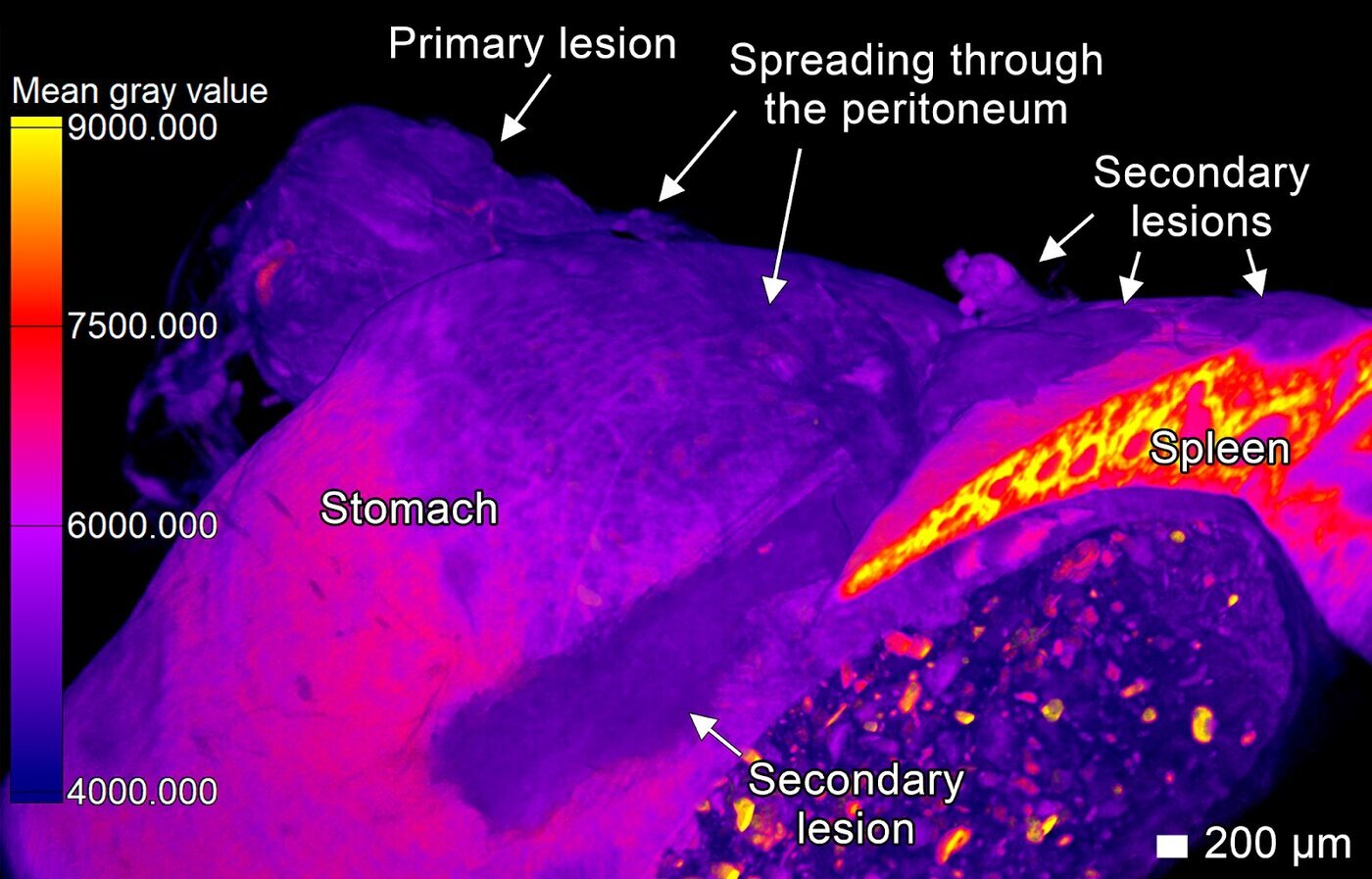
This world-first study leveraged high-resolution X-ray microtomography to capture three-dimensional visualizations of tumor progression with microscopic precision, offering previously unseen perspectives on how cancer infiltrates tissues and organs. By combining this with traditional histology, the team created a comprehensive spatial analysis of tumor behavior in living tissue.
“Our findings show that cancer is not just an aggressive disease, but a highly adaptive and coordinated system,” said Sergey Tkachev, Principal Investigator at the Institute of Regenerative Medicine at Sechenov University and lead researcher at Cancer 3D. “Metastases aren’t merely endpoints—they become active nodes, continuing the invasion in a self-sustaining loop.”
The research focused on esophageal squamous cell carcinoma, using patient-derived tumor samples transplanted into immunodeficient mice to replicate the invasive and metastatic processes. For the first time, researchers captured the collective migration of tumor cells in three dimensions. These cells exhibited complex behaviors: forming finger-like projections into the spleen, gliding across the peritoneal lining, and penetrating pancreatic tissue—all without destroying surrounding structures.
“Microtomography allowed us to reconstruct tumor spread through the peritoneum in ways that were previously only hypothesized,” said Tkachev. “We visualized how cancer cells move like coordinated units—much like social amoebas—using available tissue structures to advance with minimal destruction.”
The study provides compelling evidence for a cyclical, network-based model of metastasis, first proposed by cancer researcher Peter Friedl in 2011. It challenges the linear paradigm of tumor spread and suggests that new therapeutic strategies should focus on disrupting this self-reinforcing cycle.
Importantly, Cancer 3D’s work also revealed that traditional two-dimensional histological analysis may misinterpret critical features such as “tumor budding”—often considered a key prognostic marker. In 3D, these buds appear as extensions of the main tumor mass, not isolated structures, opening new questions about current diagnostic standards.
The implications of Cancer 3D’s work extend beyond academic insight. By enabling precise 3D imaging of surgically removed tumors, lymph nodes, and biopsy samples, this technology can significantly improve cancer staging accuracy and treatment planning. Protocols are now being developed to apply microtomography to paraffin-embedded blocks, resected breast and lung tissue, and bone biopsies.
“At Cancer 3D, our mission is to transform how the world sees cancer—literally and scientifically,” said Tkachev. “This breakthrough opens new paths for personalized medicine, especially in combating metastatic tumors where current therapies typically fail. We invite all collaborators to join the project.”
With the potential to serve as a new gold standard in tumor visualization, Cancer 3D continues to lead the charge in merging biomedical imaging, oncology, and AI-based analytics.
About Cancer 3D
Cancer 3D is a research community that uses advanced X-ray microtomography and AI-powered analytics to enable three-dimensional visualization of tumor structures and progression. Collaborating with leading research institutions and clinical centers, Cancer 3D aims to revolutionize cancer diagnostics, treatment planning, and drug development through spatial oncology. All the datasets can be found here.
Media Contact
Hanna Karasevich
Cancer 3D
Email: hannakarasevich@gmail.com
X: @cancer_3d


